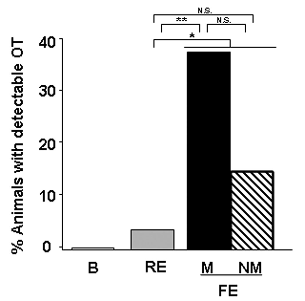
Figure 1.
In vivo microdialysis to detect extracellular oxytocin (OT) as a function of social exposure and mating. Four 30-min samples were collected and analyzed for each phase. The graph illustrates the percentage of animals yielding microdialysates in each phase with detectable OT concentrations. Under basal conditions (B) OT concentrations were below the level of detectability (<0.05 pg/sample) in all samples. Detectable OT was observed significantly more frequently during the free exposure (FE) phase compared to during the restricted exposure (RE) phase when the male was housed in a wire cage (* = Fisher’s exact test, P = 0.05). In addition, detectable OT was observed significantly more frequently during the FE phase in females that mated (M) compared to during the RE (** = Fisher’s exact test, P = 0.039). In the group of females that failed to mate (NM) during the free exposure phase, the percentage with detectable OT during that phase was not significantly different from the restricted exposure phase (Fisher’s exact test, P > 0.05). There was no significant difference in the number of females that mated vs unmated during the FE phase (Fisher’s exact test, P > 0.05).

