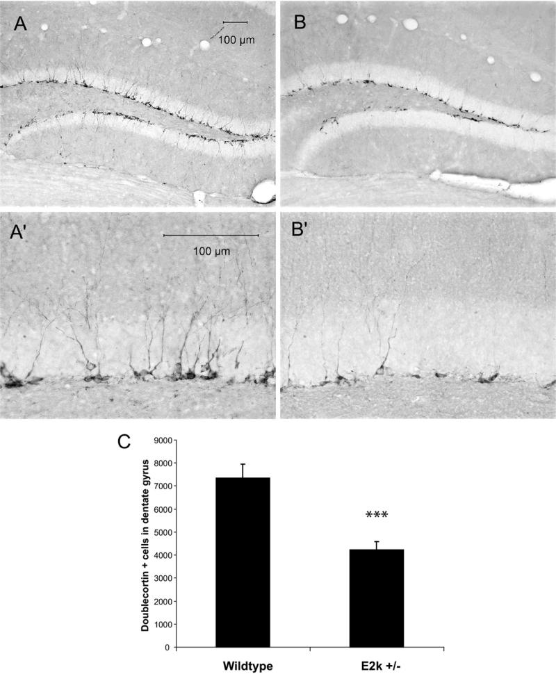
Fig. 1.
Dcx immunoreactivity in the SGZ of wild-type control (A, A′) and E2k+/– (B, B′) mice showing a reduction of Dcx-labeled cells in the latter. High magnification photos depict reductions in the number and dendritic branching of Dcx-labeled neurons in the E2k+/– mice (B′) compared with wild-type control (A′). Stereological evaluation (C) revealed a significant loss of Dcx-positive cells in the SGZ of E2k+/– mice compared with wild-type controls (*** P<0.001). The stereological cell counts represent the total number of cells in the SGZ within the specified brain region included in the analysis (see Experimental Procedures).

