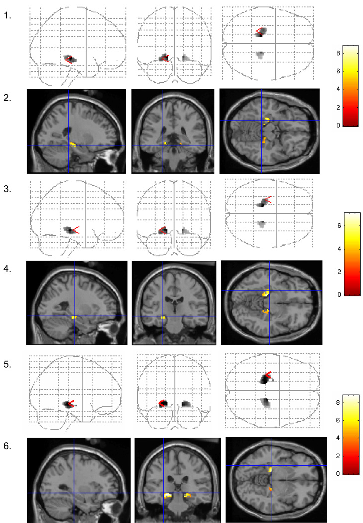
Figure 2. Characteristic activations in the medial temporal lobe during performance of verbal tasks.
Sagittal, coronal, and axial projections of medial temporal regions showing significant (p < .01) increases in activation across the two groups combined. There were no significant decreases in activation. Odd numbered rows show glass brain images of significant findings, and even numbered rows show activations superimposed on a structural anatomical template. For ease of presentation and reference to HT effects, the cross-hairs reference the regions showing HT effects in the group comparisons, as depicted in Figure 2. The scale to the right of each pair of figures shows the color scale corresponding to the z-values for that particular analysis. Rows 1 and 2 show significantly increased activation in the bilateral parahippocampal gyrus during verbal recognition across the two groups combined (in contrast to the decreased activation seen in perimenopausal HT users in Figure 1). Rows 3 and 4 show significantly increased activation across the two groups combined in the left hippocampus during verbal recognition (in comparison to the increased activation seen in perimenopausal HT users in Figure 1). Rows 4 and 5 show a similar effect during verbal match.

